Techniques
Radical Microscopes easily upgradable to techniques such as DF, DIC, Phase, IMC, Fluorescence
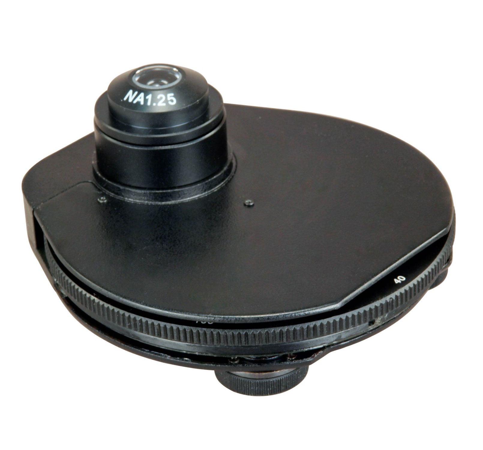
| Hoffman Modulation Contrast | Hoffman modulation contrast microscopy (IMC microscopy) is an optical microscopy technique for enhancing the contrast in unstained biological specimens. Like differential interference contrast microscopy (DIC microscopy), contrast is increased by using components in the light path which convert phase gradients in the specimen into differences in light intensity that are rendered in an image that appears three-dimensional. The 3D appearance may be misleading, as a feature which appears to cast a shadow may not necessarily have a distinct physical geometry corresponding to the shadow. The technique is particularly suitable for optical sectioning at lower magnifications Available for objectives10X, 20X & 40X with full 360° rotating modulator |
|---|---|
| DIC | Differential interference contrast DIC) microscopy, also known as Nomarski interference contrast NIC or Nomarski microscopy, is an optical microscopy technique used to enhance the contrast in unstained, transparent samples. DIC works on the principle of interferometry to gain information about the optical path length of the sample, to see otherwise invisible features. A relatively complex optical system produces an image with the object appearing black to white on a grey background. This image is similar to that obtained by phase contrast microscopy but without the bright diffraction halo. Universal condenser with turret DIC configuration for different magnification alongwith objective DIC prism having transition control are available for objective 10X, 20X, 40X, 60X & 100X. |
| Phase Contrast | Phase-contrast microscopy is an optical microscopy technique that converts phase shifts in light passing through a transparent specimen to brightness changes in the image. Phase shifts themselves are invisible, but become visible when shown as brightness variations. Universal turret (5 or 6 positions) or different Phase slider are available for 10X, 20X, 40X & 100X objective |
| Darkfield | In optical microscopy, dark-field describes an illumination technique used to enhance the contrast in unstained samples. It works by illuminating the sample with light that will not be collected by the objective lens and thus will not form part of the image. This produces the classic appearance of a dark, almost black, background with bright objects on it. Oil or dry Aplanatic Darkfield Condenser is available alongwith 100X objective having IRIS Diaphragm. Brightfield objective 4X, 10X, 20X, & 40X can be used in darkfield application alongwith darkfield condenser. Universal condenser having DF slot/position is available with other microscopy technique |
| Polarising | Polarized light microscopy can mean any of a number of optical microscopy techniques involving polarized light. Simple techniques include illumination of the sample with polarized light. Directly transmitted light can, optionally, be blocked with a polariser orientated at 90° to the illumination. More complex microscopy techniques which take advantage of polarized light include differential interference contrast microscopy and interference reflection microscopy. Scientists will often use a device called a polarizing plate to convert natural light into polarized light. With long working distance objectives specially design for polarizing application, any microscope can be upgraded to polarizing microscopy by adding polarizer and analyzer in the optical path. Specialized polarizing microscope with conoscopic observation is also available. |
| Fluorescence | A fluorescence microscope is an optical microscope that uses fluorescence and phosphorescence instead of, or in addition to, reflection and absorption to study properties of organic or inorganic substances.The "fluorescence microscope" refers to any microscope that uses fluorescence to generate an image, whether it is a more simple set up like an epifluorescence microscope, or a more complicated design such as a confocal microscope, which uses optical sectioning to get better resolution of the fluorescent image. Different type of Fluorescence illumination Mercury 100/150W illumination Xenon 75W illumination LED based illumination Alogwith fibre guided (cool) illumination is also available Basket of Fluorescence filters soured from world’s best manufacturer makes it possible to observe almost all Fluorescence dyes |